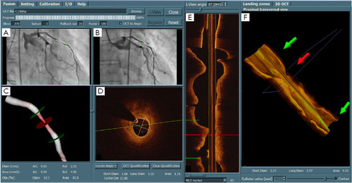
3D quantitative coronaryangiography (3D QCA) and its registration with 3D optical coherence tomography(OCT). A and B are the two angiographic views; C is the reconstructed vesselsegment in color-coded fashion; D. is the OCT cross-sectional viewcorresponding to the middle (red) marker; E is the OCT longitudinal view; and Fis the 3D OCT image. After the registration, the corresponding markers indifferent views (A, B, C, D, and F) were synchronized, allowing the assessmentof lumen dimensions from both imaging modalities at every correspondingposition along the vessel segment.
QCA, IVUS and OCT in interventional cardiology in 2011
JohanH.C. Reiber, Shengxian Tu, Joan C. Tuinenburg, Gerhard Koning, Johannes P.Janssen, Jouke Dijkstra
